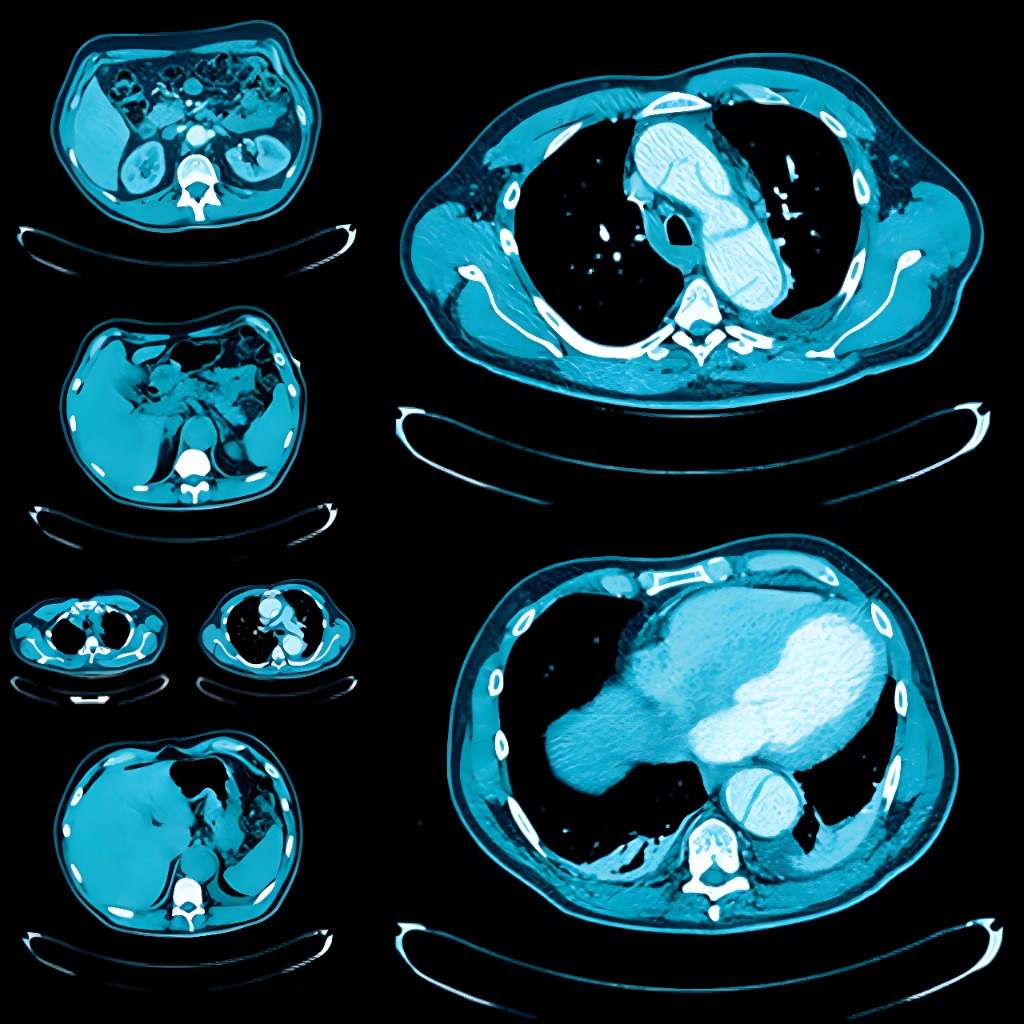
News Notification

UCSF Abdominal and Thoracic Imaging

1212 views Tags: CME UCSF Board Review

UCSF ABDOMINAL AND THORACIC IMAGING

University of California San Francisco Clinical Update (SA-CME)

This CME program details recent advances and emerging trends in abdominal and thoracic imaging, along with expert advice to optimize patient outcomes.

0 0 votes
Article Rating

Stay Current with Key Advances

UCSF Abdominal and Thoracic Imaging is an extensive review of clinically relevant topics in chest, abdominal and ob-gyn imaging. This expertly designed course includes case-based lectures on topics like imaging of neuroendocrine tumors, female pelvis and early pregnancy, acute aortic syndrome, imaging of pleural disease, imaging mimics of pelvic pathology, etc. It will help you to:

– Identify common causes of acute pelvic pain in non-pregnant females

– Recognize the appearance of ectopic pregnancy

– Diagnose common and uncommon pancreatic masses

– Illustrate the proper management of a renal incidentaloma

– Apply updated guidelines for the diagnosis and management of lung nodules, lung cancer

– Differentiate pathologic conditions from benign incidentalomas and mimics in the lungs, heart and mediastinum

CHECK SAMPLES

1. Female Pelvis and Pregnancy (8)

1

Imaging Mimics of Pelvic Pathology – Michael A. Ohliger, MD, PhD

2

MRI of Gynecologic Malignancy – Michael A. Ohliger, MD, PhD

3

Technical Tips & Tricks for MRI of the Female Pelvis – Michael A. Ohliger, MD, PhD

4

Acute Pelvic Pain in the Non-pregnant Female – Lori M. Strachowski, MD

5

Diagnosing Nonviable Pregnancy in the 1st Trimester – Lori M. Strachowski, MD

6

Ectopic Pregnancy: Rings, Rules and Role of US – Lori M. Strachowski, MD

7

O-RADS: An Introduction to the Lexicon and Risk Stratification – Lori M. Strachowski, MD

8

US of the Uterus and Endometrium: Pearls to Perfect Your US Performance – Lori M. Strachowski, MD

2. Male Pelvis and Liver (7)

1

Hepatobiliary Agents and Their Role in Liver Imaging – Thomas A. Hope, MD

2

Imaging of Biochemically Recurrent Prostate Cancer – Thomas A. Hope, MD

3

LI-RADS Cases – Thomas A. Hope, MD

4

MRI of Diffuse Liver Disease – Michael A. Ohliger, MD, PhD

5

Prostate MRI T2 and Diffusion – Michael A. Ohliger, MD, PhD

6

Rapid-body MR Imaging Techniques – Michael A. Ohliger, MD, PhD

7

Sonography of the Scrotum: A Practical Approach – Lori M. Strachowski, MD

3. Gastrointestinal CT/MR (8)

1

Imaging of Neuroendocrine Tumors – Thomas A. Hope, MD

2

Imaging of Rectal Cancer – Thomas A. Hope, MD

3

CT of Peptic Ulcer Disease – Michael A. Ohliger, MD, PhD

4

Acute Appendicitis – Emily M. Webb, MD

5

CT of Colitis: Infection, Inflammation and Ischemia – Emily M. Webb, MD

6

Cystic Pancreatic Masses: Imaging Update – Emily M. Webb, MD

7

Everything You Need to Know about the Spleen in Less than an Hour – Emily M. Webb, MD

8

Primary Retroperitoneal Masses: An Approach to Diagnosis – Emily M. Webb, MD

4. Abdominal and Thoracic (8)

1

Acute Aortic Syndrome – Travis S. Henry, MD

2

Imaging of Pleural Disease – Travis S. Henry, MD

3

Mediastinal Masses – Travis S. Henry, MD

4

How to Handle Thoracic Incidentalomas – Kimberly G. Kallianos, MD

5

Look at the Heart: Cardiac Findings on Non-gated CT – Kimberly G. Kallianos, MD

6

Update on Pulmonary Embolism – Kimberly G. Kallianos, MD

7

Acute Abdominal Pain – Emily M. Webb, MD

8

Interesting Cases in the Abdomen and Pelvis – Emily M. Webb, MD

5. Thoracic and Pulmonary

1

Bronchiectasis: An Imaging Approach – Travis S. Henry, MD

2

Introduction to HRCT – Travis S. Henry, MD

3

Lateral Chest Radiograph – Travis S. Henry, MD

4

Lung Nodules: What Do I Ignore, and What Do I Further Explore? – Travis S. Henry, MD

5

Small Airways Disease – Travis S. Henry, MD

6

Challenging CXR Cases – Kimberly G. Kallianos, MD

7

Confidently Diagnosing Lung Cancer – Kimberly G. Kallianos, MD

8

Pulmonary Infections – Kimberly G. Kallianos, MD

Learning Objectives

Upon completion of this course, you will know how to:

– Diagnose common causes of acute pelvic pain in non-pregnant women

– Recognize the appearance of ectopic pregnancy

– Utilize ultrasound to identify findings suggestive and definitive of pregnancy failure in the first trimester

– Differentiate common solitary liver masses

– Describe the proper management of a renal incidentaloma

– Evaluate the small and large bowel in patients with abdominal pain

– Diagnose common and atypical pancreatic masses

– Apply updated guidelines for the diagnosis and management of lung nodules and lung cancer

– Recognize typical and atypical appearance of thoracic emergencies including pulmonary embolism and acute aortic syndromes

– Differentiate pathologic conditions from benign incidentalomas and mimics in the lungs, heart and mediastinum

– Apply standard terminology to improve description and classification of interstitial lung disease, small airways disease and bronchiectasis on HCRT

Intended Audience

This educational activity was designed for radiologists and other medical professionals who would benefit from a greater understanding of the interpretation of abdominal and thoracic images.

Designation

Series Release: April 1, 2019

Series Expiration: March 31, 2022

Subscribe
Notify of
guest
0 Comments
Inline Feedbacks
View all comments
error: Content is protected !!