News Notification

NYU’s Head to Toe Imaging 2020

1107 views Tags: CME Oakstone Board Review

NYU's HEAD TO TOE IMAGING 2020

NYU Grossman School of Medicine Clinical Update (SA-CME)

Take an informative look at key radiology topics with special focus on recent advances and evolving trends in imaging technology, disease diagnosis and management across all sub specialty areas.

0 0 votes
Article Rating

Stay Ahead in Your Field

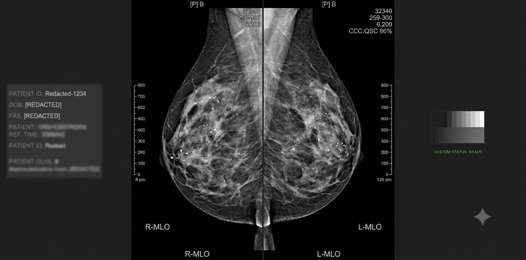
NYU’s Head to Toe Imaging is extremely thorough and will help you keep up with recent developments and current trends in the field. Led by Georgeann McGuinness, MD, FACR, this online CME program focuses on safety and quality issues, the practical aspects of established CT, MR, US, PET, and interventional techniques, and new techniques and applications for these modalities. It will help you to better:

– Evaluate and fit current imaging techniques and protocols for sub specialty imaging into clinical practice

– Explain the Bosniak classification system for small renal masses

– Develop imaging protocols to optimize radiation safety and dose reduction in CT

– Utilize MR imaging in apt patient populations without compromising diagnostic accuracy

– Strategize to optimize and incorporate a gamut of studies, including CT, MR and PET to accurately stage neoplasms

CHECK SAMPLES

1. Neuroradiology (9)

1

Pediatric Brain Tumors: What is New – Alireza Radmanesh, MD

2

Adult Brain Tumors: Radiogenomics – Rajan Jain, MD

3

Stroke Imaging – Seena Dehkharghani, MD

4

Adult Acquired White Matter Disorders – Benjamin Cohen, MD

5

The Irvin I. Kricheff Lecture – Metastatic Neck Disease: Assessment of Unknown Primary – Vijay M. Rao, MD, FACR

6

Sinuses: Inflammatory Diseases and Post Operative Imaging – Vijay M. Rao, MD, FACR

7

Transverse Myelitis – Maria J. Borja, MD

8

AJCC Update in HN Cancer Staging – Elcin Zan, MD

9

Post-operative Surveillance in HN Tumors – Mari Hagiwara, MD

2. Interventional Radiology (3)

1

Prostate Artery Embolization: From Diagnostic Workup to Post-Procedure Imaging – Cash Horn, MD

2

Imaging and Management of Portal Hypertension – Bedros Taslakian, MD

3

LI-RADS and Interventional Oncology Follow-Up – Eric T. Aaltonen, MD, MPH

3. Interventional and Neuroradiology Challenging Cases (5)

1

Challenging IR Cases – Meredith R. McDermott, MD

2

Neuroradiology ED Cases – Joseph Boonsiri, MD

3

Non-Vascular – Brain and Spine Cases – Anna Derman, MD

4

Vascular – Brain and Spine Cases – Matthew G. Young, DO

5

Head and Neck Cases – Gopi K. Nayak, MD

Lorem ipsum dolor sit amet...

1

Imaging of the Postoperative Breast – Alana A. Lewin, MD

2

Tips and Tricks for Sonographic Correlation – Chloe Chhor, MD

3

Mammographic Analysis of Breast Calcifications – Edward A. Sickles, MD

4

Diagnostic Evaluation with Tomosynthesis – Beatriu Reig, MD, MPH

5

Mammographic Follow-Up as an Alternative to Biopsy: Strengths, Limitations, and Pitfalls – Edward A. Sickles, MD

6

Hormones, Imaging Biomarkers, and Breast Cancer Risk – Samantha L. Heller, PhD, MD

7

A New Perspective on Male Breast Cancer – Yiming Gao, MD

8

Updates on Fast Breast MRI and Advanced Breast MRI Techniques – Laura Heacock, MD

9

Medicolegal Aspects of Breast Imaging – Edward A. Sickles, MD

Lorem ipsum dolor sit amet...

1

Pediatric Hip – Mark E. Bittman, MD

2

Pediatric Knee – Lynne P. Pinkney, MD

3

Pediatric Elbow – Shailee Lala, MD

Lorem ipsum dolor sit amet...

1

Pediatric Cases – Netta M. Blitman, MD

2

Breast Imaging of the Young, Pregnant, and Lactating Patient – Elizabeth V. Mausner, MD

3

Challenging Image-guided Breast Procedure Cases – Kristin R. Elias, MD

Lorem ipsum dolor sit amet...

1

Shoulder Imaging: Pearls and Pitfalls – Soterios Gyftopoulos, MD, MSc

2

Elbow Imaging: Pearls and Pitfalls – William R. Walter, MD

3

Groin Injuries – Erin Alaia, MD

4

Hip Cartilage and Labral Injuries: What the Surgeon Wants to Know – Donna G. Blankenbaker, MD, FACR

5

Knee Avulsion Injuries – prepared by Zehava S. Rosenberg, MD, presented by Mohammad M. Samim, MD

6

AI in MSK Imaging – Michael P. Recht, MD

7

MSK Infections: How to Avoid Misdiagnosis – Donna G. Blankenbaker, MD, FACR

8

Surgery of the Lumbar Spine: The Basics – Leon D. Rybak, MD

9

Weird Bone Lesions – Mohammad M. Samim, MD

10

Ultrasound of Lumps and Bumps – Ronald. S. Adler, MD, PhD

11

Ultrasound Guided Procedures – Christopher Burke, MD

Lorem ipsum dolor sit amet...

1

PET Imaging of Colorectal Carcinoma – Munir Ghesani, MD, FACNM, FACR

2

PET Imaging of Prostate Cancer – Shawn Karls, MD

3

Management of Neuroendocrine Tumors with Ga-Dotatate PET – Elcin Zan, MD

Lorem ipsum dolor sit amet...

1

MSK Cases: Upper Extremity – Renata La Rocca Vieira, MD

2

PET Results and Path Reports: Concordance or “Learning Opportunity?” – Munir Ghesani, MD, FACNM, FACR

3

MSK Cases: Lower Extremity – Michael B. Mechlin, MD

4

Ga-Dotatate PET: False-positives and Incidental Malignancies – Elcin Zan, MD

Lorem ipsum dolor sit amet...

1

Central Airway Diseases – Maria C. Shiau, MD

2

An Imaging Approach to Small Airway Disease – Travis S. Henry, MD

3

Seeing the Picture in a Mosaic – Georgeann McGuinness, MD, FACR

4

Thoracic Sarcoidosis: Hidden in Plain View – Jeffrey B. Alpert, MD

5

Shades of Gray: Subsolid Lung Nodules – Lea Azour, MD

6

Leveling the Playing Field: Navigation Guided Thoracic Intervention – William H. Moore, MD

7

State of the Art: CT Pulmonary Angiography – Jane P. Ko, MD

8

Smoke: Differentiating Flow Artifact from Pathology on Thoracic CTA – Travis S. Henry, MD

Lorem ipsum dolor sit amet...

1

Wrong Way Turns: Coronary Anomaly Misses and Misperceptions – Lea Azour, MD

2

Mama Told Me There’d Be Days Like This: Challenging Cardiac CT Cases – Jill E. Jacobs, MD, FACR, FAHA

3

Extraordinary Cases in Congenital Heart Disease – Dan Halpern, MD

Lorem ipsum dolor sit amet...

1

Challenging Chest Cases: To Err is Human – Francis G. Girvin, MD

2

Segmental Approach to Congenital Heart Disease – Puneet Bhatla, MD

3

Zebras in the Chest Reading Room – Geraldine Brusca-Augello, DO

4

Cardiovascular MRI Cases: What We Got Wrong and Why – Leon Axel, PhD, MD

Lorem ipsum dolor sit amet...

1

Practicing Green CT – Alec J. Megibow, MD, MPH, FACR

2

Ultrasound Diagnosis of Non-gynecologic Causes of Pelvic Pain – Genevieve L. Bennett, MD

3

Small Bowel Imaging – Kira Melamud, MD

4

The Morton A. Bosniak Lecture – Imaging of Solid Renal Masses: Lessons Learned from Bosniak Classification – Hersh Chandarana, MD, MBA

5

Prostate MR – Andrew B. Rosenkrantz, MD

6

Bariatric Imaging Pearls from the Practicing Surgeon – Manish S. Parikh, MD

7

Top 10 Updates in Body Imaging from 2019 – Nicole M. Hindman, MD

8

Challenging Benign Focal Liver Lesions – Nicole M. Hindman, MD

9

Fibrosis, Cirrhosis, and HCC: An Imaging Update – Krishna Shanbhogue, MD

10

Post Y90 Liver Imaging – Sooah Kim, MD

11

Rectal MRI – Myles Taffel, MD

Lorem ipsum dolor sit amet...

1

Mass Casualty Incidents: An Introduction for Imagers – Mark P. Bernstein, MD, FASER

2

Facial Trauma – Sergey Kochkine, MD

3

Emergency Pelvic US – Aspan S. Ohson, MD

Lorem ipsum dolor sit amet...

1

Interesting and Unusual ER Cases – Alexander Baxter, MD

2

TI-RADS – Chrystia Slywotzky, MD

3

Renal Cases – Stella Kang, MD

4

Vascular Cases – ChenChan Huang, MD

5

Bowel Cases – Bari Dane, MD

6

Gynecologic Cases – Angela Tong, MD

7

Body ER Cases – Jay Karajgikar, MD

Learning Objectives

After viewing this program, the participant will be better able to:

– Evaluate and incorporate current imaging techniques and protocols for sub specialty imaging (Abdominal, Musculoskeletal, Neurologic, Thoracic, Cardiac, Breast, PET/CT, Emergency Medicine, Interventional) into clinical practice to enable accurate diagnosis, dictate best therapy options, and assess response to therapy, which may prompt therapy modification as needed.

– Describe the Bosniak classification system, and its applicability to watchful waiting/surveillance management algorithms for small renal mass lesions.

– Describe the recent literature and recommendations for imaging lung nodules and imaging appearances of various subsolid nodules.

– Develop strategies to optimize and incorporate a gamut of studies, including CT, MR and PET, in order to accurately stage neoplasms with minimal cost and radiation exposure.

– Develop imaging protocols and techniques to optimize radiation safety and dose reduction in CT, as well as utilize MR imaging in appropriate patient populations, without compromise to diagnostic quality and accuracy of imaging.

Intended Audience

The program is designed for clinical radiologists in either general or specialized practice, and is appropriate for radiologists in training. Familiarity with the fundamentals of CT, MR, PET, and US technology and applications, as well as basic anatomy, is assumed.

Designation

Date of Original Release: February 15, 2020

Date of Original Release: February 15, 2023

Subscribe
Notify of
guest
0 Comments
Oldest
Newest Most Voted
Inline Feedbacks
View all comments
error: Content is protected !!