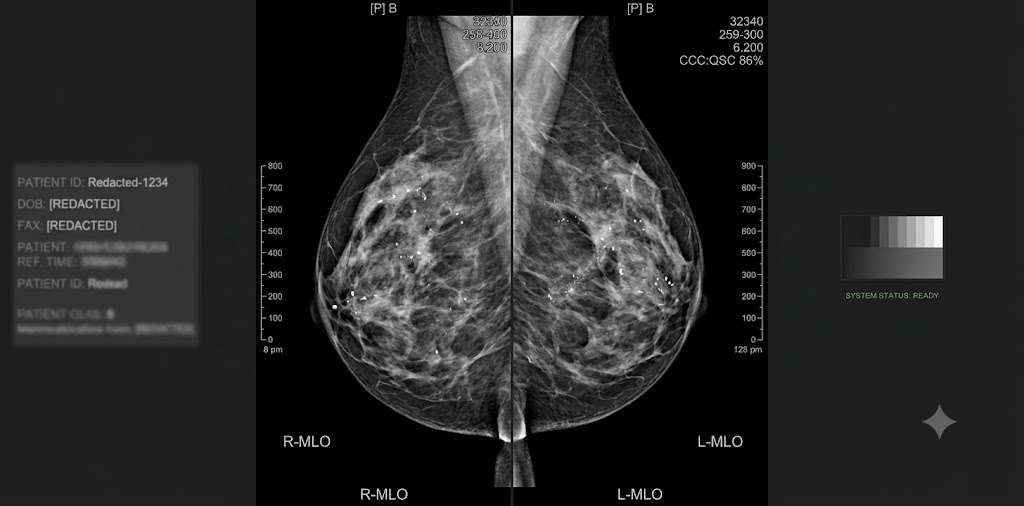
Stay Current on Rapidly Changing Specialty
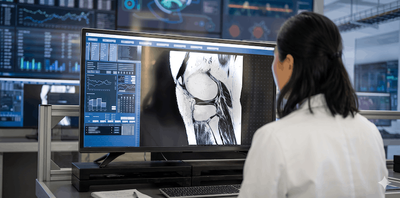
UCSF Neuro and Musculoskeletal Imaging is a comprehensive state-of-the-art update on clinically relevant topics in neuroradiology and musculoskeletal imaging. This in-depth CME course includes case-based lectures on topics like skeletal trauma, shoulder instability, brain tumor pitfalls, hydrocephalus, staging cervical nodes, and more. It will help you to better:
- Revamp imaging protocols for brain, head & neck, spine, nerve and musculoskeletal imaging
- Understand the parameters of MR arthrographic technique and interpretation of findings
- Develop strategies to create short brain, spine and neck differentials
- Implement newer MR sequences like 3D volumetric imaging and metal suppression techniques
- Understand advances in stroke management and its effect on imaging
- Outline strategies to evaluate common and uncommon abnormalities in the neck and spine
* Date of Original Release: April 16, 2019
* Date of Expiration: April 15, 2022
Learning Objectives
Upon completion of this activity, participants will be able to apply the following:
- Update and improve imaging protocols for brain, head & neck, spine, nerve and musculoskeletal imaging
- Recognize specific imaging features of infection and tumors in the head, neck, spine and peripheral nerves
- Distinguish between normal anatomy, common anatomic variants and pathological disorders related to MRI of the major musculoskeletal joints, brain, head, neck and spine
- Recognize internal derangement appearances of the knee, shoulder, elbow, wrist, hip, knee, and foot
- Implement newer MR sequences such as 3D volumetric imaging and metal suppression techniques
- Understand the parameters of MR arthrographic technique and interpretation of findings
- Sharpen evaluation of muscle and tendon abnormalities, and evaluate various abnormalities that simulate musculoskeletal tumors
- Develop strategies for creating short brain, spine and neck differentials
- Understand advances in stroke management and its impact on imaging
- Develop strategies for evaluating common and uncommon abnormalities in the neck and spine
UCSF NEURO AND MUSCULOSKELETAL IMAGING
UCSF NEURO AND MUSCULOSKELETAL IMAGING TOPICS
CHECK SAMPLES
1. Hydrocephalus – A. James Barkovich, MD
2. Imaging of Neuro Phakomatoses – A. James Barkovich, MD
3. Imaging of Normal and Injured Neonatal and Infant Brain – A. James Barkovich, MD
4. Infections of the Pediatric Brain – A. James Barkovich, MD
5. Techniques for Neonatal Imaging without Sedation – A. James Barkovich, MD
6. ACL Reconstruction – Matthew D. Bucknor, MD
7. MRI of Hip Impingement – Matthew D. Bucknor, MD
8. Osseous Infectious Dilemmas – Matthew D. Bucknor, MD
9. Radiographic Checklist for Hip Impingement – Matthew D. Bucknor, MD
10. The Throwing Elbow – Matthew D. Bucknor, MD
11. Brain Tumor Pitfalls – William P. Dillon, MD
12. CNS Infections – William P. Dillon, MD
13. Current Concepts in Stroke Imaging – William P. Dillon, MD
14. Imaging of Headache – William P. Dillon, MD
15. White Matter Beyond Multiple Sclerosis – William P. Dillon, MD
16. Head and Neck Cases – 1 – Christine M. Glastonbury, MBBS
17. Head and Neck Cases – 2 – Christine M. Glastonbury, MBBS
18. Neck Infections – Christine M. Glastonbury, MBBS
19. Parotid Masses – Christine M. Glastonbury, MBBS
20. Staging Cervical Nodes – Christine M. Glastonbury, MBBS
21. Unknown Primary Tumors – Christine M. Glastonbury, MBBS
22. CNS Hypotension: Finding and Fixing that Elusive Leak – Vinil N. Shah, MD
23. CNS Spine Emergencies: Top 5 Diagnoses Not-to-Miss – Vinil N. Shah, MD
24. Practical Brachial Plexus MRI – Vinil N. Shah, MD
25. Rapid-Fire Neuro Cases – Vinil N. Shah, MD
26. Value-Based Imaging of the Degenerative Spine – Vinil N. Shah, MD
27. Ankle MRI: Tendons and Ligaments – Ramya Srinivasan, MD
28. Hip Ultrasound Made Easy – Ramya Srinivasan, MD
29. Knee Ligaments – Ramya Srinivasan, MD
30. Skeletal Trauma: Commonly Missed Injuries – Ramya Srinivasan, MD
31. Knee Menisci: Pearls and Pitfalls – Lynne S. Steinbach, MD
32. MRI of the Post-Operative Shoulder – Lynne S. Steinbach, MD
33. Pearls and Pitfalls in Shoulder MRI – Lynne S. Steinbach, MD
34. Shoulder Instability – Lynne S. Steinbach, MD
35. The Throwing Shoulder – Lynne S. Steinbach, MD