News Notification

National Diagnostic Imaging Symposium 2020

907 views Tags: CME

NATIONAL DIAGNOSTIC IMAGING SYMPOSIUM™ 2020

World Class CME Clinical Update

Expand your medical knowledge and diagnostic skills with expert presentations covering 10 radiology subspecialties.

0 0 votes
Article Rating

Learn from the Experts

World Class CME’s National Diagnostic Imaging Symposium™ is a comprehensive program featuring 115 presentations across 10 subspecialties, including Abdominal, Chest, Cardiovascular, Emergency, Genitourinary, Neurology, Musculoskeletal and Nuclear Medicine. This CME course will help you to better:

– Assess injuries and diseases of the extremities, spine and face using CT and MRI

– Apply ultrasound in musculoskeletal, gynecological, obstetrical, cardiovascular and other organ systems

– Use MDCT and MR imaging to evaluate the abdomen, liver, pancreas, spleen and bowel

– Evaluate genitourinary malignancies, masses and anomalies using MRI, CT and US

– Understand the newest breast screening and diagnostic technologies

– And more…

CHECK SAMPLES

1. Chest Radiology (11)

1

Imaging of Fibrotic Interstitial Lung Disease: The New Classification of UIP and the Rationale for Change – Jonathan H. Chung, MD

2

Smoking-related Lung Disease – Caroline Chiles, MD

3

Imaging of Cystic Lung Disease – Thomas E. Hartman, MD, FACR

4

Airways Disease: A Case-based Approach – Jared D. Christensen, MD

5

Lung Nodule Management Guidelines: What to Use When – Jared D. Christensen, MD

6

Lung Cancer Screening: A Case-based Review – Caroline Chiles, MD

7

Diffuse Nodular Lung Disease – Jonathan H. Chung, MD

8

Pulmonary Complications in the Immunocompromised Host – Thomas E. Hartman, MD, FACR

9

Incidental Findings on Chest CT – Caroline Chiles, MD

10

Occupational Lung Disease – Jonathan H. Chung, MD

11

Imaging Evaluation of Acute Chest Pain – Jared D. Christensen, MD

2. Musculoskeletal Radiology (5)

1

The Wrist: Intrinsic Ligaments and Tendons – Mark W. Anderson, MD

2

The Shoulder: Labral Pathology – Laura W. Bancroft, MD, FACR

3

MRI of the Elbow: A Case-based Review – Mark W. Anderson, MD

4

Imaging of Osteoporosis and its Complications – Mark W. Anderson, MD

5

The Knee: Ligaments – Laura W. Bancroft, MD, FACR

6

Imaging of Lateral Ankle Pain – Laura W. Bancroft, MD, FACR

3. Breast MRI (11)

1

Point-Counter-Point: Abbreviated Screening Breast MRI Versus Breast MRI – Keep the Full Protocol – Sarah M. Friedewald, MD, and Bonnie N. Joe, MD, PhD

2

3T vs 1.5T in Breast: What Are the Benefits? Are They Needed? – Katja Pinker, MD, PhD, EBBI

3

Controversies in Breast MRI – Are They Real? – Christopher E. Comstock, MD, FACR

4

MRI Biopsies: From Basics to Tips and Tricks – Sarah M. Friedewald, MD

5

Breast MRI in the Post Lumpectomy Patient – Christopher E. Comstock, MD, FACR

6

The Effective Use of Breast MRI as a Problem Solver – Katja Pinker, MD, PhD, EBBI

7

Current Status of Breast DWI – Bonnie N. Joe, MD, PhD

8

Breast MRI in the Preoperative Setting – Sarah M. Friedewald, MD

9

Challenges with BIRADS-3 in Breast MRI – Christopher E. Comstock, MD, FACR

10

Difficulties with NME: Is it BPE or DCIS? – Katja Pinker, MD, PhD, EBBI

11

Breast MRI Cases to Leave For Your Partner – Bonnie N. Joe, MD, PhD

4. Gastrointestinal Radiology (12)

1

MR for the Non-Traumatic Acute Abdomen – Perry J. Pickhardt, MD

2

Acute Pancreatitis – Koenraad J. Mortele, MD

3

Demystifying Peritoneal Disease – Perry J. Pickhardt, MD

4

Solid Pancreatic Tumors Beyond Adenocarcinoma – Koenraad J. Mortele, MD

5

MDCT Evaluation of the Spleen – Elliot K. Fishman, MD

6

Evaluation of Bowel Ischemia – Richard M. Gore, MD

7

CT of Small Bowel Tumors – Elliot K. Fishman, MD

8

Appendicitis and Diverticulitis and Their Mimics – Richard M. Gore, MD

9

Understanding NAFLD – Perry J. Pickhardt, MD

10

MRC of Benign Biliary Disorders – Koenraad J. Mortele, MD

11

MDCT of Parenchymal Liver Disease – Elliot K. Fishman, MD

12

Diagnosis and Staging of Biliary Neoplasms – Richard M. Gore, MD

5. Ultrasound (12)

1

Why You Should Be Offering Liver Elastography in Your Practice – Richard G. Barr, MD, PhD

2

There is a Mass in the Scrotum: What Does it Mean? – Thomas C. Winter III, MD

3

Sonography of the First Trimester – Peter Doubilet, MD, PhD

4

Pediatric Ultrasound from the ER: Right Lower Quadrant Pain: More Than Appendicitis – Marilyn J. Siegel, MD

5

How Contrast Enhanced Ultrasound Will Improve Your Patient Outcomes – How to Get Started – Richard G. Barr, MD, PhD

6

Medical Malpractice Issues in Obstetrics for the General Radiologist – Thomas C. Winter III, MD

7

Ectopic Pregnancy: Diagnosis – Peter Doubilet, MD, PhD

8

Pediatric Ultrasound from the ER: Imaging Turns and Twists of Bowel Obstruction – Marilyn J. Siegel, MD

9

Contrast Enhanced Ultrasound of the Liver and Kidney – Richard G. Barr, MD, PhD

10

Interesting Abdominal Ultrasound Cases – Thomas C. Winter III, MD

11

Placenta, Cord, and Amniotic Fluid – Peter Doubilet, MD, PhD

12

Pediatric Ultrasound From the ER: Acute Scrotum: Practice Essentials – Marilyn J. Siegel, MD

6. Breast Imaging (12)

1

The Community-based Breast Imaging Center – How to Make it Efficient and Profitable – Stamatia Destounis, MD, FACR

2

High Risk Screening: The Devil is in the Details – Maxine S. Jochelson, MD

3

Medicolegal Issues in Breast Imaging – Edward A. Sickles, MD

4

How to Reduce the Frequency of Missed Cancers – Edward A. Sickles, MD

5

Case Studies of Potential Lawsuits – What Can We Learn? – Edward A. Sickles, MD

6

Ultrasound Features of Breast Masses as Biomarkers – A. Thomas Stavros, MD, FACR

7

Tomosynthesis in the Screening and Diagnosis Environments – Stamatia Destounis, MD, FACR

8

Contrast Enhanced Mammography: State of the Art – Maxine S. Jochelson, MD

9

Breast Imaging in Women Receiving Neoadjuvant Chemotherapy – Maxine S. Jochelson, MD

10

Challenging Cases in Tomosynthesis, What Can We Learn? – Stamatia Destounis, MD, FACR

11

Ultrasound of Nipple Discharge and Papillary Lesions – A. Thomas Stavros, MD, FACR

12

An Analysis of Ultrasound False Negative Findings – A. Thomas Stavros, MD, FACR

7. Emergency Radiology (12)

1

MDCT Maxillofacial Trauma: Saving Face – Mark P. Bernstein, MD

2

Shoulder Trauma – Manickam Kumaravel, MD

3

Pitfalls in CT of Blunt Abdominal Trauma – Jorge A. Soto, MD

4

CT and MR of Spine Infections – Wayne S. Kubal, MD

5

Musculoskeletal Emergencies – Manickam Kumaravel, MD

6

Using MR to Address Problematic Cervical Injuries – Wayne S. Kubal, MD

7

Easily Missed Thoracolumbar Injuries – Mark P. Bernstein, MD

8

CT and MR of Biliary Tract Emergencies – Jorge A. Soto, MD

9

Injuries to the Aorta and Diaphragm – Mark P. Bernstein, MD

10

Imaging of Crohn’s Disease in the ER – Jorge A. Soto, MD

11

Avoiding Errors: Foot and Ankle – Manickam Kumaravel, MD

12

Non-Traumatic Subarachnoid Hemorrhage – Wayne S. Kubal, MD

8. Genitourinary (12)

1

Imaging of Adrenal Masses – Stuart G. Silverman, MD, FACR

2

Imaging of Treated and Recurrent Prostate Cancer – Hebert Alberto Vargas, MD

3

MRI of the Bladder and Urethra – Mukesh Harisinghani, MD

4

MRI of Adnexal Lesions – Iva Petkovska, MD

5

Renal Mass Imaging Update – Stuart G. Silverman, MD, FACR

6

Imaging of Cervical and Endometrial Malignancies – Hebert Alberto Vargas, MD

7

Prostate MR: PIRADS 2 Simplified – Mukesh Harisinghani, MD

8

MRI of Benign Conditions of the Uterus – Iva Petkovska, MD

9

Case-based Review: Pitfalls of GU MRI – Hebert Alberto Vargas, MD

10

Case-based Review: GU Infections – Mukesh Harisinghani, MD

11

Case-based Review: MRI of Acute Pelvic Pain in Women – Iva Petkovska, MD

12

Case-based Review: CT Urography – Stuart G. Silverman, MD, FACR

9. Advanced Topics in Breast (7)

1

A Revolutionary New Era in the Diagnosis and Treatment of Breast Cancer, Past, Present and Future – László K. Tabár, MD

2

Modern and Effective Calcification Analysis – László K. Tabár, MD

3

Architectural Distortion on the Mammogram – Detection and Workup Algorithm Leading to the Correct Diagnosis – László K. Tabár, MD

4

Tomosynthesis – It Just Keeps Getting Better! – Elizabeth A. Rafferty, MD

5

Current Screening Guidelines: One Step Forward, Two Steps Back? – Elizabeth A. Rafferty, MD

6

Am I Missing Something? Minimizing False Positives with Tomosynthesis – Elizabeth A. Rafferty, MD

7

Through the Looking-Glass: A Potpourri of Case Studies – Elizabeth A. Rafferty, MD

10. Neuroradiology (12)

1

Sellar and Parasellar Lesions – James G. Smirniotopoulos, MD

2

Spine Neoplasms – Erik Gaensler, MD

3

Stroke Intervention: What Really Matters in Your Reports – James E. Lefler, MD

4

Brain Infections – Blake A. Johnson, MD, FACR

5

Neuroimaging of Drug Abuse – Camilla E. Lindan, MD

6

Tumor Markers in Diffuse Gliomas – James G. Smirniotopoulos, MD

7

Head and Neck Tumors – Blake A. Johnson, MD, FACR

8

AVM’s A-Z – Erik Gaensler, MD

9

Upping Your Game! From Common Indications to Stellar Report: Headache – Camilla E. Lindan, MD

10

Upping Your Game! From Common Indications to Stellar Report: Cognitive Decline – Camilla E. Lindan, MD

11

Intracranial Hemorrhage – Blake A. Johnson, MD, FACR

12

Mysteries of Metastases – James G. Smirniotopoulos, MD

11. Cardiovascular Radiology (8)

1

Pearls and Pitfalls for Carotid Duplex Interpretation – John S. Pellerito, MD, FACR

2

CT of the Abdominal Aorta: Aneurysms, Dissections, and Repair – Elliot K. Fishman, MD

3

Multimodality Imaging in the Assessment of Structural Heart Disease – Michael Poon, MD, FACC

4

Doppler Evaluation of Portal Hypertension – John S. Pellerito, MD, FACR

5

CT of GI Bleeding – Elliot K. Fishman, MD

6

Noninvasive Evaluation of Chest Pain – Michael Poon, MD, FACC

7

CTA of the Abdominal Vessels and Runoff Studies – Elliot K. Fishman, MD

8

Doppler Evaluation of Renal and Mesenteric Arteries – John S. Pellerito, MD, FACR

Learning Objectives

At the conclusion of this activity, the participant will be able to:

– Increase diagnostic capabilities in multiple radiology sub-specialties

– Utilize CT and MRI to assess injury and disease in the extremities, spine and face

– Apply ultrasound to musculoskeletal, gynecological, obstetrical, cardiovascular and other organ systems

– Effectively use MDCT and MR imaging to evaluate the abdomen, liver, pancreas, spleen and bowel

– Plan the appropriate imaging of emergency department patients, including pediatric patients

– Improve assessment of genitourinary malignancies, masses and anomalies using MRI, CT and US

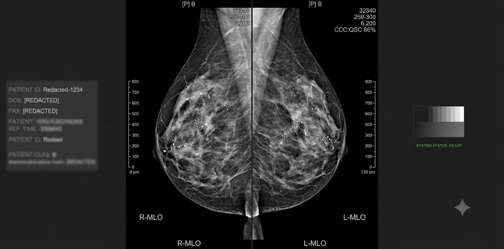
– Improve diagnostic accuracy in breast imaging and distinguish advantages of the newest breast screening and diagnostic technologies

– Correctly interpret and stage CT findings in the context of a lung cancer-screening program

– Understand how to utilize non-invasive options such as CT, MR and ultrasound for the evaluation of vascular problems

– Improve skills in MR and US-guided radiology procedures

– Enhance understanding of the use and limitations of PET/CT

Intended Audience

This program was designed to meet the educational needs of practicing radiologists, radiologists in training and other physicians, as well as advanced practitioners with a special interest in radiology.

Designation

Date of Original Release: February 15, 2020

Date of Original Release: February 15, 2023

Subscribe
Notify of
guest
0 Comments
Inline Feedbacks
View all comments
error: Content is protected !!