An Acclaimed Review of Radiology Fundamentals
The UCSF Radiology Review – Comprehensive Imaging is appropriate for residents, clinical fellows and practicing radiologists interested in updating their working knowledge, by covering all the major radiology subspecialties. The course represents the breadth and depth of experience from each subspecialty while providing a step-by-step process for reaching a differential diagnosis.
This CME program will help you to:
– Identify imaging fundamentals of each of the radiology sub-specialties and major organ systems
– Prescribe the appropriate utilization of imaging modalities (CT, MR, US, Mammography, Nuclear Medicine) for different medical conditions
– Implement the appropriate imaging protocols for routine clinical presentations
– Recognize indications and markers for interventional procedures, as well as assessment of outcomes
– Apply classifications of pathophysiological mechanisms underlying human disease to facilitate differential diagnosis
– Refine imaging criteria to optimize image quality/findings and improve diagnostic interpretation
CHECK SAMPLES
1. Gastrointestinal
|
1
|
Pancreatic Tumors and Mimics – Emily M. Webb, MD |
|
2
|
CT/MR of the Acute Abdomen – Derek Sun, MD |
|
3
|
Imaging of Colon Cancer Emily – M. Webb, MD |
|
4
|
Biliary Tract Benjamin – M. Yeh, MD |
|
5
|
Liver DiseaseThomas – A. Hope, MD |
|
6
|
Fluoroscopy – Spencer C. Behr, MD |
2. Genitourinary
|
1
|
Benign Gynecologic Disease – Liina Poder, MD |
|
2
|
Malignant Gynecologic DiseaseMichael A. Ohliger, MD, PhD |
|
3
|
Non-Tumorous Renal Disease – Priyanka Jha, MBBS |
|
4
|
Renal & Adrenal Masses – Ronald J. Zagoria, MD, FACR |
|
5
|
Bladder, Prostate, Urethra – Ronald J. Zagoria, MD, FACR |
3. Thoracic, Pulmonary
|
1
|
HRCT of the Lung: Findings-Based Approach to Diagnosis – Brett M. Elicker, MD |
|
2
|
CT/PET of Pulmonary Nodules – David M. Naeger, MD |
|
3
|
The Lateral Chest Radiograph – Travis S. Henry, MD |
|
4
|
Lung Cancer Staging – David M. Naeger, MD |
|
5
|
CT/PET of Mediastinal Tumors – David M. Naeger, MD |
4. Cardiac, Physics, Safety
|
1
|
Imaging of the Heart: Top 10 Diagnoses – Kimberly G. Kallianos, MD |
|
2
|
Vascular Diseases of the Thorax – Michael D. Hope, MD |
|
3
|
Practical Physics for the Radiologist: CT – Michael A. Ohliger, MD, PhD |
|
4
|
Practical Physics for the Radiologist: MR – Michael A. Ohliger, MD, PhD |
|
5
|
Ultrasound Technical Tips/Artifacts – John T. Mongan, MD, PhD |
|
6
|
IV Contrast Use Update – Ronald J. Zagoria, MD, FACR |
|
7
|
Playing it Safe: What You Need to Know About CT/MR Safety – John T. Mongan, MD, PhD |
5. Pediatrics
|
1
|
The Building Blocks of Pediatric Musculoskeletal Imaging – John D. MacKenzie, MD |
|
2
|
Imaging the Pediatric Acute Abdomen: Pearls & Pitfalls – Jesse Courtier, MD |
|
3
|
Pediatric Scrotal Imaging: The “Ts” – Jesse Courtier, MD |
|
4
|
Pediatric Chest Imaging: What Not-to-Miss – Matthew Zapala, MD, PhD |
|
5
|
Updates in Non-Accidental Trauma – Matthew Zapala, MD, PhD |
|
6
|
Important Pediatric Abdominal Masses – Andrew S. Phelps, MD |
6. Nuclear Medicine, Interventional
|
1
|
When PET Has Low Sensitivity – David M. Naeger, MD |
|
2
|
FDG PET Artifacts and Incidentalomas – Spencer C. Behr, MD |
|
3
|
Vascular Diagnosis and Interventions – K. Pallav Kolli, MD |
|
4
|
GI/GU Interventions – Nicholas Fidelman, MD |
|
5
|
Percutaneous Biopsies – K. Pallav Kolli, MD |
|
6
|
Portal and Biliary Interventions – Maureen P. Kohi, MD |
|
7
|
Interventional Oncology – Nicholas Fidelman, MD |
|
8
|
Interventions in Women’s Imaging – Maureen P. Kohi, MD |
7. Ultrasound/OB-GYN
|
1
|
Abdominal Ultrasound – Tara A. Morgan, MD |
|
2
|
Key Points for Thyroid Imaging and Biopsy – Tara A. Morgan, MD |
|
3
|
Female Pelvis: Uterus/Adnexa – Liina Poder, MD |
|
4
|
Key Points in the First Trimester – Liina Poder, MD |
|
5
|
Key Points in the Second Trimester – Ruth B. Goldstein, MD |
|
6
|
Most Relevant Fetal Anomalies – Vickie A. Feldstein, MD |
|
7
|
Vascular Ultrasound – Dorothy J. Shum, MD |
|
8
|
Genitourinary Ultrasound – Priyanka Jha, MBBS |
8. Breast
|
1
|
Masses and Asymmetries – Bonnie N. Joe, MD, PhD |
|
2
|
Calcifications – Heather I. Greenwood, MD |
|
3
|
Breast MRI – Bonnie N. Joe, MD, PhD |
|
4
|
Radiology-Pathology Concordance – Amie Y. Lee, MD |
|
5
|
Breast Ultrasound – Jessica H. Hayward, MD |
9. Neuro
|
1
|
Brain Tumors – Soonmee Cha, MD |
|
2
|
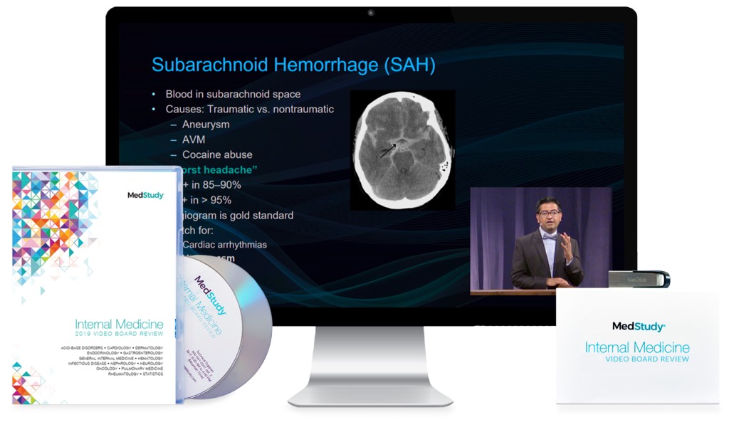
Intracranial Hemorrhage – Esther L. Yuh, MD, PhD |
|
3
|
Brain Infections – Jason F. Talbott, MD, PhD |
|
4
|
Spine Masses – Vinil N. Shah, MD |
|
5
|
Head & Neck Masses – Alina Uzelac, DO |
10. Musculoskeletal
|
1
|
Shoulder MRI – Rina Patel, MD |
|
2
|
Knee MRI – Ramya Srinivasan, MD |
|
3
|
Arthritis and Infection – Thomas M. Link, MD, PhD |
|
4
|
Skeletal Trauma: Extremities – Terry C.P. Lynch, MD |
|
5
|
Metabolic – Robert D. Boutin, MD |
|
6
|
Tumors & Tumor-like Conditions – Robert D. Boutin, MD |
Learning Objectives
After viewing this activity, participants will demonstrate the ability to:
– Identify imaging fundamentals of each of the radiology sub-specialties (organ systems)
– Prescribe the appropriate utilization of imaging modalities (CT, MR, US, Mammography, Nuc Med) for different medical conditions
– Implement the appropriate imaging protocols for routine clinical presentations
– Recognize indications and markers for interventional procedures, as well as assessment of outcomes
– Apply classifications of pathophysiological mechanisms underlying human disease to facilitate differential diagnosis
– Refine imaging criteria to optimize image quality/findings and improve diagnostic interpretation
Intended Audience
This comprehensive radiology overview is appropriate for residents, clinical fellows and practicing radiologists interested in updating their working knowledge, of all the major radiology subspecialties.
Designation
Series Release: July 1, 2018
Series Expiration: June 30, 2021